Technical File on Thermography
Infrared Thermography System?
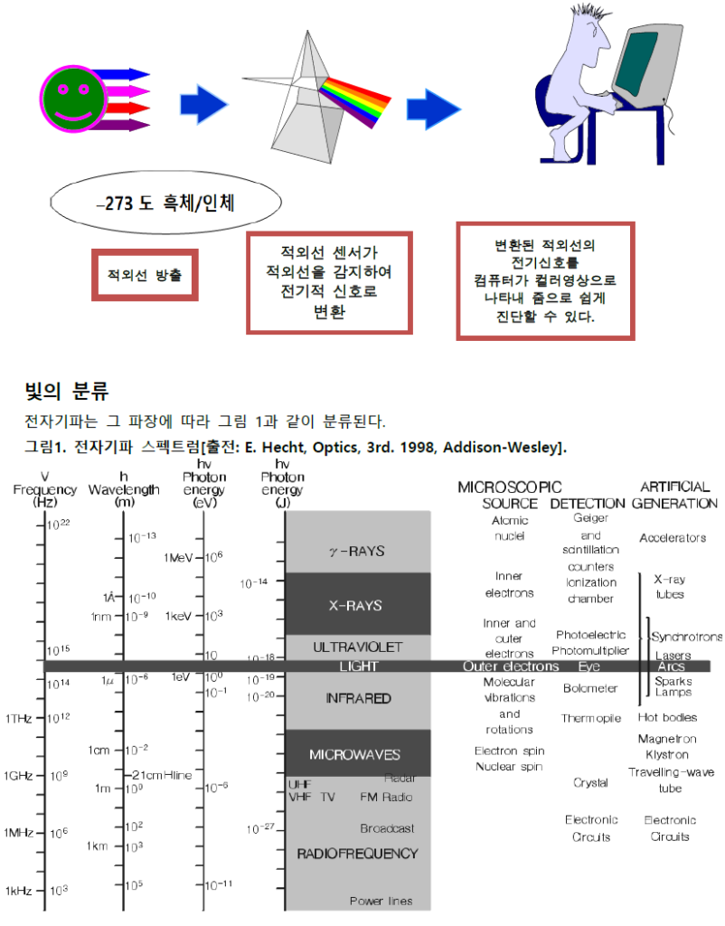
Infrared Thermography System displays the thermal images of sensitive changes on the pain area by detecting the extra minimum infrared ray emitting from the body.
It is very useful equipment to diagnose the Neuropathy, Breast cancer, Physiological Problems, Inflammation and Circulatory disorder which are difficult to be found by CT or MRI without any radiation.
Also, it can be used on pregnant women and infants since it’s non-invasive and harmless to the human body.
1. Principle of Thermography System
Infrared thermography imaging systems work based on the principle of detecting the infrared radiation emitted by objects.
All objects warmer than absolute zero(0 Kelvin or -273.15℃) emit energy somewhere within above range.
Infrared thermography is obtaining this heat energy from subjects and transforming into digital, colored image using highly sophisticated equipment including infrared camera, electrical signal converter, and computer.
Infrared camera detects the 8 to 12 µm infrared range emitted from the human body and displays areas where a lot of infrared energy is emitted as high temperatures and areas where less infrared energy is emitted as low temperatures.
Here’s how it works :
1. Detection of Infrared Radiation: Infrared cameras contain sensors that are sensitive to infrared radiation. These sensors can detect the intensity of the radiation emitted by objects.
2. Conversion to Temperature: The intensity of the infrared radiation detected by the camera is converted into temperature values. This conversion is based on the relationship between an object’s temperature and the amount of infrared radiation it emits.
3. Image Formation: The converted temperature values are then used to generate an image. In this image, different colors or shades represent different temperatures. Warmer areas are usually depicted in warmer colors like red or orange, while cooler areas appear as cooler colors like blue or green.
4. Visualization and Analysis: The resulting thermal image can be analyzed to identify temperature variations across the scene. This information can be useful in various applications such as detecting heat leaks, monitoring equipment for signs of overheating, and identifying anomalies in structures or processes.
2. Relationship between Body Temperature and Skin
The relationship between body temperature and the skin is multifaceted and crucial for maintaining thermal equilibrium and overall health. Here are some key points regarding this relationship:
Thermoregulation: The skin plays a central role in thermoregulation, the process by which the body maintains its core temperature within a narrow range. When the body temperature rises above normal, such as during physical exertion or exposure to heat, blood vessels in the skin dilate (vasodilation), allowing more blood to flow near the skin’s surface. This facilitates heat dissipation through radiation, conduction, and convection, helping to cool the body.
Temperature Sensation: The skin contains thermoreceptors, specialized nerve endings that detect changes in temperature. These receptors provide feedback to the brain about the external temperature environment and the temperature of the skin itself. This information helps the body adjust its thermoregulatory responses accordingly.
Evaporative Cooling: Sweat glands in the skin produce sweat, which evaporates from the skin’s surface, carrying away heat and helping to cool the body. This process is particularly important during periods of elevated body temperature, such as during exercise or in hot environments.
Fever Response: During fever, which is often a response to inflammation or illness, the body’s core temperature is elevated. The skin may feel warm to the touch as blood vessels dilate to facilitate heat loss.
The influence of the external environment appears as an important variable in measuring skin temperature.
Drugs or cosmetics act as a barrier, making measurement impossible, and even affect clothing, humidity, temperature, and air flow around the subject.
Heat enters and exits through the skin, but about 25% is conducted through conduction, 25% is through evaporation, and only about 45% escapes to the outside through radiation.
In addition, because radiation is done on the surface, there is a big difference depending on the posture of the person being measured, and each part of the human body also varies depending on its anatomical structure.
Factors that affect blood circulation, such as drinking and smoking, should also be controlled.
At temperatures below 17℃, blood vessel constriction occurs, and at temperatures above 25℃, evaporation due to sweating occurs, so measurements should be conducted at a temperature 21~25℃.
3. Diagnostic Criteria for Thermography
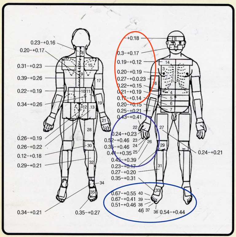
Our body temperature is controlled by the temperature control center located in the hypothalamus to maintain homeostasis. Therefore, in a healthy subject, there is a contralateral (right and left) symmetry in the skin temperature distribution, and an asymmetry above a certain level is a strong indicator of an abnormality.
Therefore, the most basic diagnosis is when the temperature distribution on both sides is asymmetrical thermal pattern, which is considered abnormal.
In a study of body temperatures in a series of healthy subject, the overall average temperature difference is within 0.3℃. The temperature difference differs in each part of the body and the various guidelines have been recommended for diagnosis of thermal asymmetry.
Generally, the mean temperature difference greater than 0.5℃ is considered to be abnormal. When greater than 1.0℃, there is no doubt of functional abnormality.
(Cranial-Caudal relationship, which shows normal temperature difference of upper and lower body, also can be used for diagnosis of bilateral disease.)
3. Concept of Real & Relative Temperature in Medical Thermography
All thermal images produced by infrared camera have the characteristic that the measured temperature varies depending on changes in the environment.
Medical thermography is a diagnostic imaging procedure produces an image of a patient’s skin surface temperature using infrared radiation, and the infrared camera commonly used in thermography uses a relative temperature measurement method in which the measured temperature changes when the environment changes.
On the other hand, the real temperature measurement method can accurately measure it even if the environment changes.
Medicore provides thermal image data using the actual temperature measurement method based on our own temperature compensation algorithm and camera manufacturing technology.
In addition, we are continuously conducting research and development so that our real temperature measurement technology can be used for Ai, big data, and thermal data standardization.