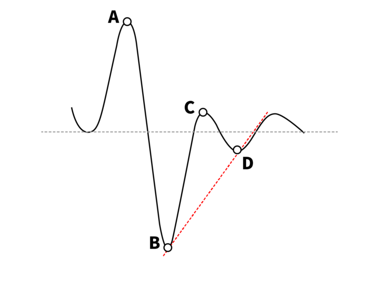
1. What is APG (Accelerated Photoplethysmograph?) Accelerated photo plethysmograph (APG) uses the second derivative of the waveform of the phtoplethysmography to stabilize the baseline and to separate components of the waveform more clearly than the first derivative. Using APG, arterial vessel elasticity, peripheral vessel elasticity, and aging index of blood vessel can be analyzed. 1) Photoplethysmograph – When blood is released from the aorta due to the heart contraction, the volume changes in blood vessels are indicated as a waveform. 2) Accelerated Photoplethysmograph – The second derivative of the waveform of the phtoplethysmography that can be used for the aging of blood vessels 2. APG Wavefrom Analysis A: Basic point to evaluate APG waveform B: Initial Systolic wave indicating the intensity of cardiac output [Arterial Elasticity] C: Late systolic re-increased wave D: Late systolic re decreased wave [Peripheral Elasticity]
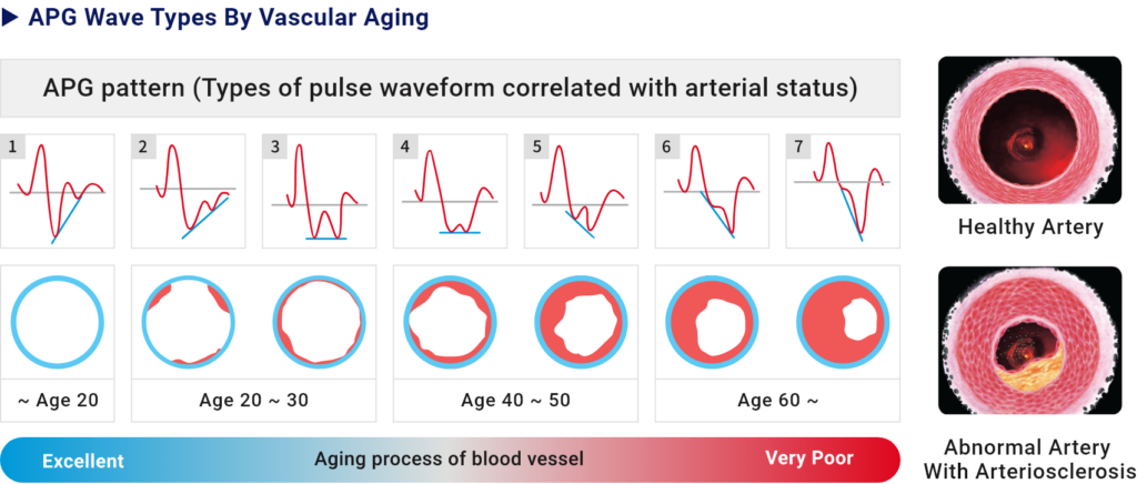
Type 1 : Blood circulation and vessel status are Excellent Type 2 : Blood circulation and vessel status are good Type 3~4 : Blood vessel starts to get aged Type 5 : Blood circulation is not good due to the aged blood vessel Type 6 : Blood circulation disorder